The quadruple helix: a new epigenetic marker?
We all know what DNA looks like, or do we? We take a look at why the quadruple helix can occur and how it could unlock new avenues for cancer therapy
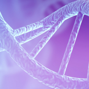
One of the things we all imagine when we think about DNA is the double helix. The iconic shape, and the race to discover it, made American biologist James Watson and English physicist Francis Crick household names.
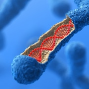
We now know, however, that DNA does not always appear in that form: sometimes it has four strands instead of two.
From double to quadruple
DNA is composed of nucleotides, which each contain one of four bases: adenine (A), cytosine (C), guanine (G) and thymine (T). These bases pair with each other – A with T, C with G – and it is the bonds between these pairs that hold the two complementary strands of DNA together in the double helix.
Guanine is unique among the bases, however, as it can also bond with itself, and when four guanine bases form a square, a four-stranded DNA structure can result – a quadruple helix.
Discovery in cells
These four-stranded DNA structures are known as G-quadruplexes, or G4s. They had been observed in the laboratory in 1988 but evidence that they were present in cells was not published until 2013.
The 2013 paper showed that G4s are formed in a range of different human cancer cells. The researchers were able to show that the structures were present, and to count them at different stages in the cell cycle. The numbers varied, indicating that G4s are transitory and most numerous when the cell’s DNA is replicated, just prior to cell division.
The team also observed that G4s tend to form in both the telomeres (important regulatory regions at the ends of each chromosome) as well as in the body of chromosomes. This fits with the observation that regulatory regions of the genome are more likely to contain sequences with a high proportion of guanine bases, which would be necessary for G4s to form.
The researchers used a chemical to ‘fix’ quadruple helices whenever they form inside the cell – this was what allowed them to be counted, but also disrupted normal cellular interactions with the G4s.
A recent paper develops the research, tagging G4s in a way that allows them to be observed within a functioning living cell. This approach has been used to demonstrate that G4s are formed in healthy cells by normal cellular processes.
The findings add to evidence that viewing DNA as synonymous with the double helix may be oversimplified and outdated.
An epigenetic function?
The reason that G4s form is still unknown, but the authors of the 2020 paper suggest that the structures form temporarily to hold the double helix open, allowing transcription to take place and therefore influencing which genes are expressed. In this way, G4s seemingly have an epigenetic effect, and may be considered a new class of epigenetic marker.
This interpretation would appear consistent with the observation that G4s are much more prevalent in cancer cells than in healthy cell types, and that the regulatory regions associated with G4 formation are also associated with cancer-causing variants.
The researchers are hopeful that an improved understanding of G4s may open up new targets for cancer therapies.
–


