Mitosis
Mitosis is a type of cell division that occurs in almost all our cells and is necessary for our bodies to grow, develop and repair. During mitosis, two identical daughter cells are produced from a single cell.
From one cell to many
Preceding mitosis, as with meiosis, each chromosome is replicated to form sister chromatids joined at the centromere (figure 1a).
As the cell enters mitosis, the chromosomes become attached by their centromeres to the spindle (figure 1b). The sister chromatids are then pulled apart and dragged to opposite sides of the cell (figure 1c). This means there is now a full complement of 46 chromosomes at each side of the cell.
The cell membrane then restricts in the middle, splitting the cell in two (figure 1d). The result: two identical diploid daughter cells (figure 1e).
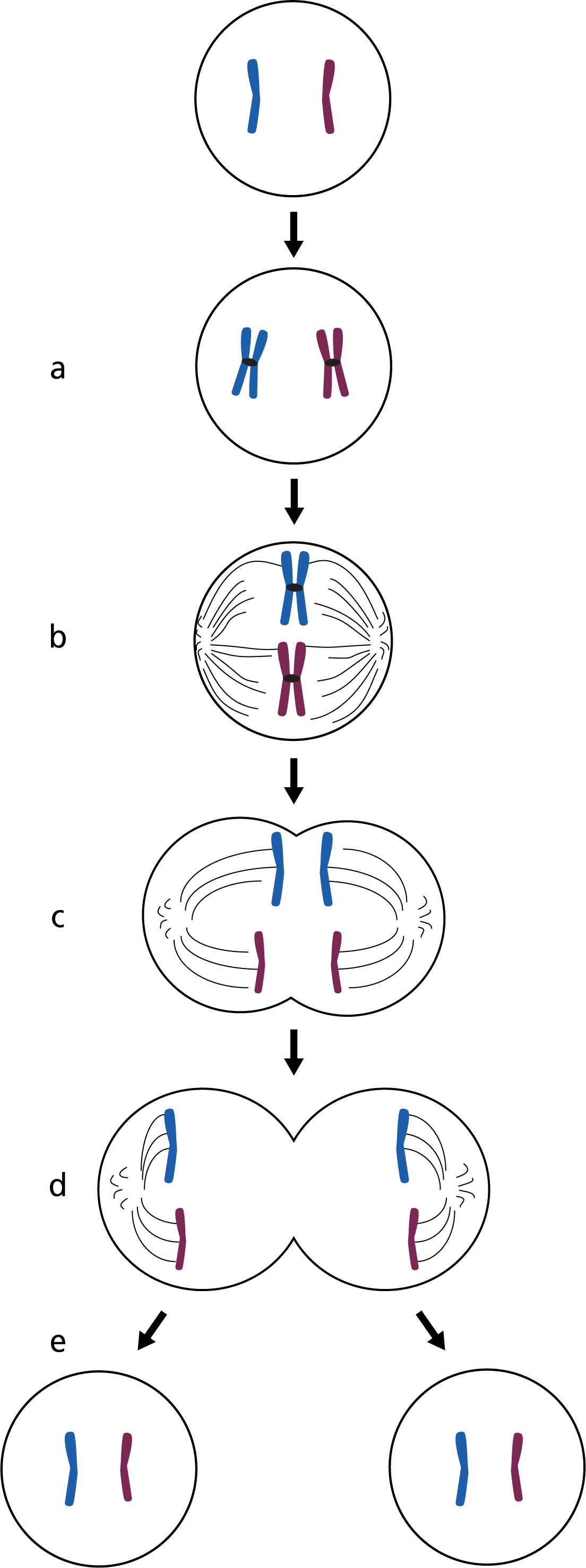
Figure 1: The stages of mitosis.
- Chromosome duplication forming sister chromatids.
- Chromosomes line up in the centre of the cell attached to the mitotic spindle.
- The spindle contracts and the sister chromatids are pulled apart to opposite ends of the cell.
- The cell constricts and divides in two.
- Two identical diploid daughter cells are produced.
Even though mitosis is essential following fertilisation, it doesn’t only occur during these early stages of development. This type of cell division not only allows the embryo and baby to grow, but it continues throughout life to repair the body or replace cells that have died.
Unlike meiosis, the function of mitosis is to produce two identical daughter cells with exactly the same complement of chromosomes.
How we inherit genomic information
Watch the following video for a summary of both meiosis and mitosis:
Key messages
- Mitosis is a type of cell division that produces two identical daughter cells from a single diploid cell.
- Mitosis occurs in nearly every cell in the human body.
- It is essential for our body’s growth, development and repair.
Resources
For clinicians
- NHS England’s Genomics Education Programme: Genomics 101: Inheriting Genomic Information