Meiosis
Meiosis is a type of cell division that only occurs in the testes and the ovaries, and forms specialised cells known as gametes. It is fundamental in the passing of genomic information from parent to offspring, while also creating genomic diversity in future generations.
Replicating the chromosomes
Meiosis begins in a specific cell called a germ cell, which like most of the cells in our bodies is diploid, with 46 chromosomes in 23 pairs.
Before meiosis begins, the DNA in each chromosome is replicated. The two identical copies that result from this DNA replication are referred to as ‘sister chromatids’, and are joined to each other by a region of the chromosome called the centromere (as shown in figure 1).
What follows are two steps of cell division, called meiosis I and meiosis II.
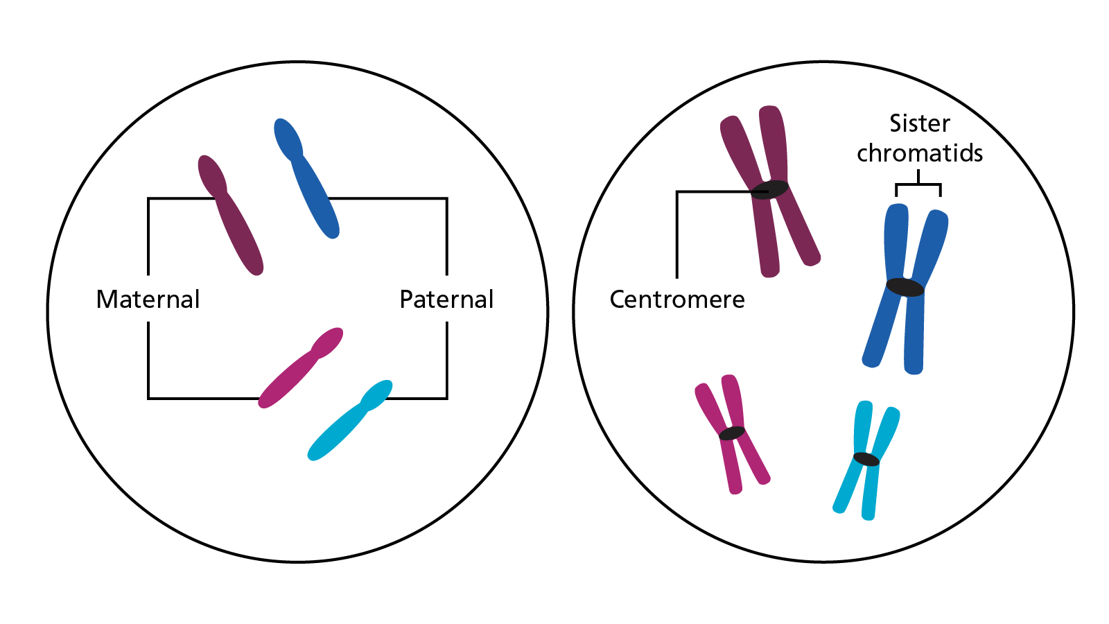 Figure 1: Two pairs of chromosomes before they are replicated (left) and after DNA replication (right) before meiosis begins
Meiosis I
Recombination
During meiosis I, the chromosomes align with their homologues (figure 2a). At this point, the homologous chromosomes break, overlap, and recombine; swapping sections of DNA between the non-sister chromatids in a process called crossing over or recombination (figure 2b).
This recombination serves to produce chromosomes with unique combinations of alleles. The maternal chromosomes will now have sections of DNA from the paternal chromosomes and vice versa (figure 2c). For the group of organisms called eukaryotes, which includes humans, there is typically one to four crossover events per homologous pair per meiosis.
Recombination
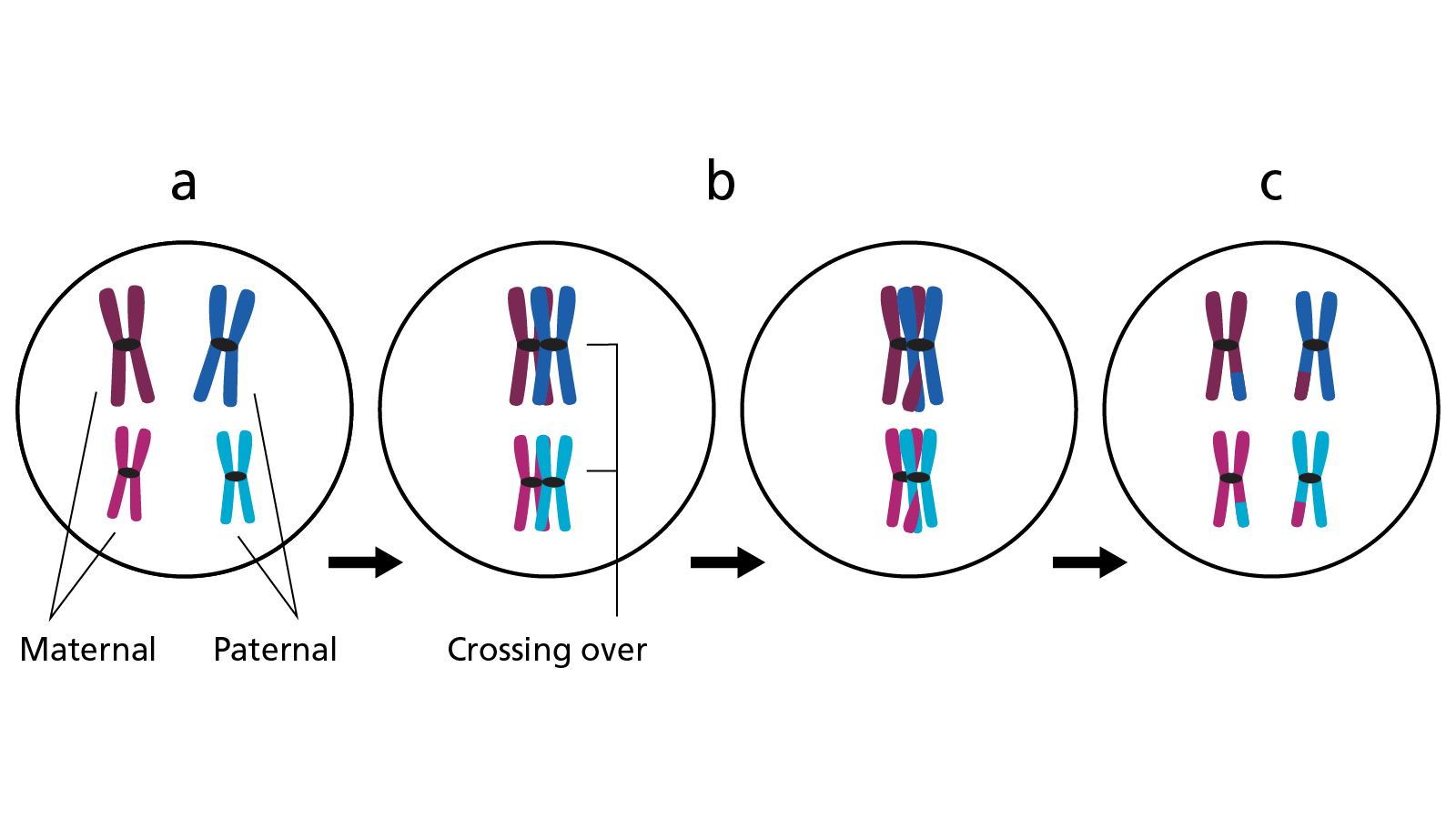 Figure 2: The stages of recombination
- Homologous chromosomes align.
- DNA is swapped between sister chromatids – recombination.
- The resulting maternal chromosomes will now have sections of DNA from the paternal chromosomes and vice versa.
Chromosome capture
The chromosomes are then captured, via their centromeres, by the meiotic spindle, a protein structure that forms during cell division (see figure 3). The fibres of the spindle span the cell like a washing line and serve to equally separate chromosomes before a cell divides. Once attached to the spindle, the chromosomes move to the middle of the cell and the homologous pairs are arranged in such a way that half are on one side and half on the other.
Chromosome capture
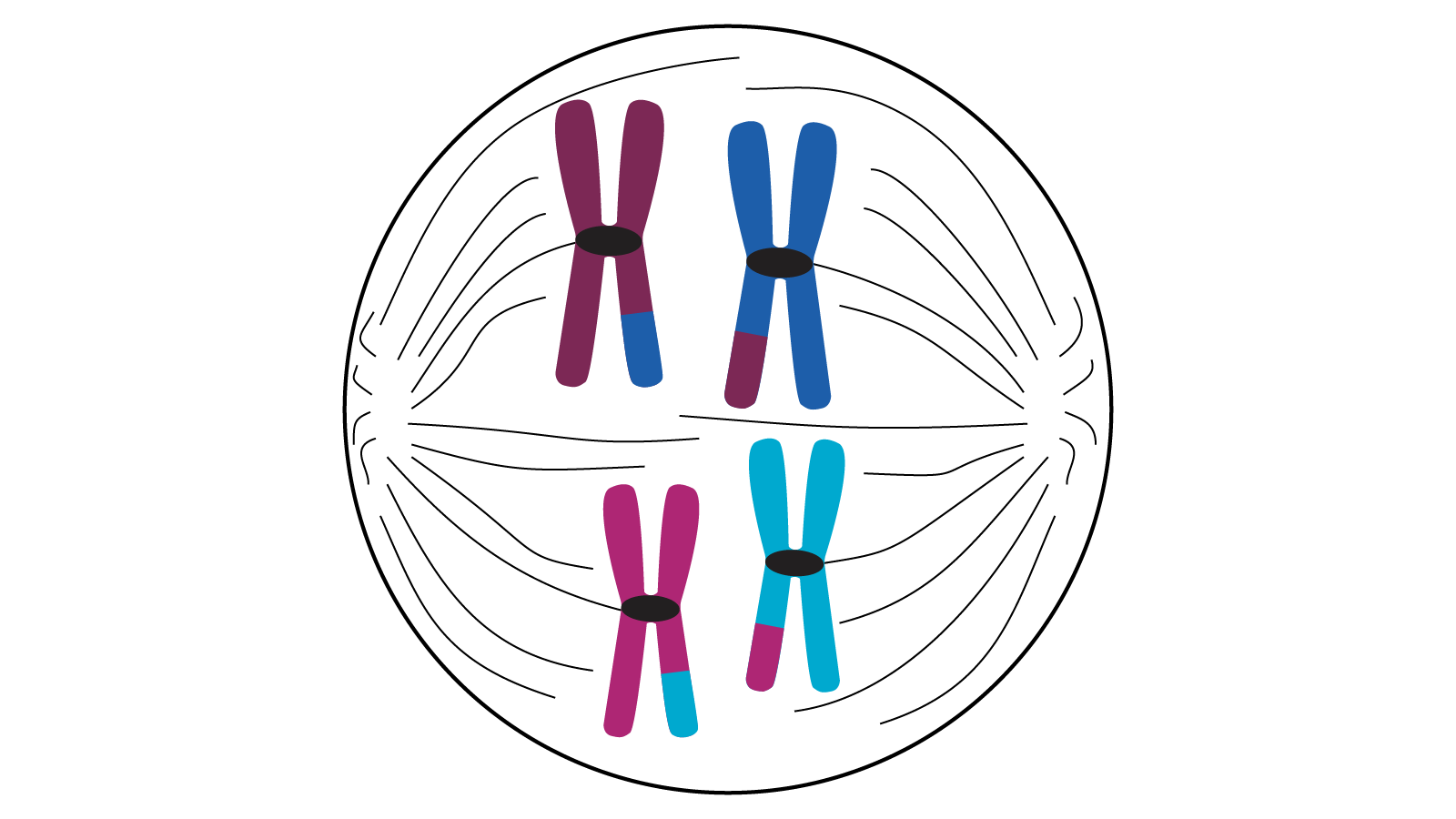 Figure 3: Four chromosomes lined up in the centre of a cell attached to the meiotic spindle
Random assortment
How the chromosomes are arranged at the middle of the cell is random, which is an additional way to make sure that the maternal and paternal genomic material is ‘mixed up’. This is known as random assortment. Owing to the number of chromosomes in the cell, there are millions of different ways this can happen. To simplify, one pair would give two possible orientations, but as humans have 23 pairs, this equates to more than 8 million possibilities.
Figure 4 shows how two pairs of chromosomes can be arranged to produce four permutations.
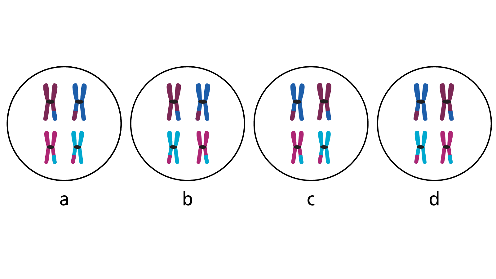 Figure 4: Two pairs of chromosomes arranged to produce four permutations
Daughter cells
The spindle then contracts and the chromosome pairs are pulled away from each other to opposite sides of the cell (Figure 5a). Due to their orientation, which side each half of the pair ends up at will be random. This results in a mixture of maternal and paternal chromosomes at either side of the cell.
At this point the cell constricts in the middle, splitting the cell in two (Figure 5b). Two new daughter cells known as primary gametes are produced, containing one chromosome from each homologous pair, but still joined to its sister chromatid (Figure 5c).
Daughter cells
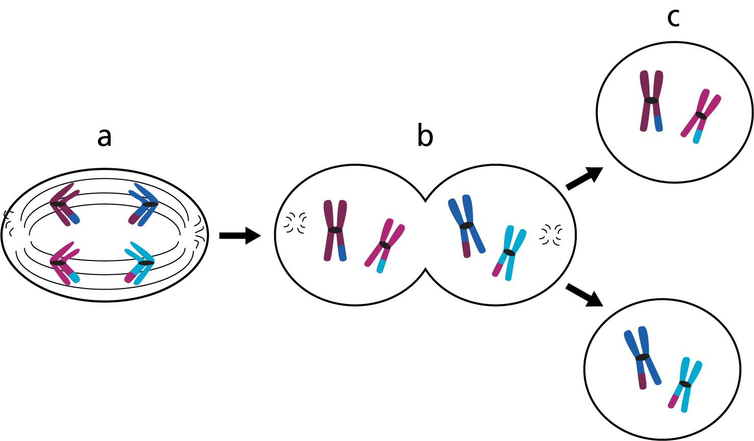 Figure 5: The cell splits in two
- Chromosome pairs are pulled to opposite sides of the cell.
- The cell constricts in the middle.
- Two primary gametes are produced, containing one chromosome from each homologous pair, but still joined to its sister chromatid.
Meiosis II
The next round of cell division, meiosis II, now begins. Unlike meiosis I, no replication of the chromosomes occurs. Instead the spindle forms and captures the chromosomes, which line up individually at the cell’s centre (Figure 6a). When the spindle contracts the sister chromatids are pulled apart to opposite ends of the cell (Figure 6b). The daughter cells constrict and divide (Figure 6c), producing four new cells called secondary gametes (Figure 6d).
Each of the secondary gametes contains one chromatid from each sister (now called a chromosome), totalling 23 in each. Gametes are haploid, as they contain half the number of chromosomes from the original diploid germ cell. In other words, the germ cell contained two sets (46) of chromosomes and the secondary gametes contain only one set (23).
Thus, at the end of meiosis, four gametes with a haploid genome have been produced from one diploid germ cell.
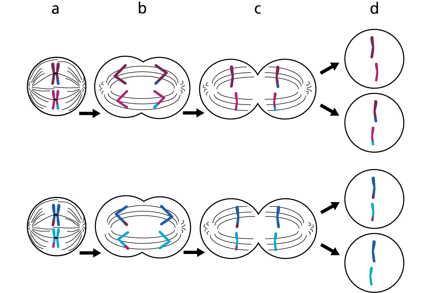 Figure 6: The stages of meiosis
- Spindle forms and captures the chromosomes, which line up individually at the cell’s centre.
- The spindle contracts and the sister chromatids are pulled apart to opposite ends of the cell.
- The daughter cells constrict and divide.
- Four new cells called secondary gametes are produced.
Differences between the sexes in meiosis
The formation of a sperm and egg is similar, although there are some differences.
For instance, the production of sperm, called spermatogenesis, begins at the onset of puberty and follows the processes described above, resulting in four gametes each time.
The production of eggs, called oogenesis, on the other hand begins when the individual is still in the womb, but then stops at the early stages of meiosis I. Once puberty is reached, meiosis continues. However, due to the different way the cells divide during oogenesis, only one functioning gamete is produced per meiosis.
Fertilisation
The cell that forms from the fusion of the sperm and egg is called a zygote. The zygote contains a full complement of chromosomes, and it is from this cell that the embryo develops and grows. For this to occur, the zygote must undergo a different type of cell division called mitosis.
How we inherit genomic information
Watch the following video for a summary of both meiosis and mitosis.
Key messages
- Meiosis is a type of cell division by which gametes – eggs and sperm – are formed.
- It includes two rounds of cell division to produce four haploid cells from a single diploid cell.
- Several processes during meiosis ensure the genomic material is ‘mixed up’ to ensure the resulting zygote contains a unique genome.
- At the point of fertilisation, the haploid sperm and egg fuse to form a diploid zygote.
Resources
For clinicians
- NHS England’s Genomics Education Programme: Genomics 101: Inheriting Genomic Information
References:
- Crismani W and Mercier R. ‘What limits meiotic crossovers?‘. Cell Cycle 2012: volume 11, issue 19, pages 3,527–3,528. DOI: 10.4161/cc.21963