Amelogenesis imperfecta
Amelogenesis imperfecta is a rare genetic condition affecting the formation of dental enamel. It can result in aesthetic and functional issues, as well as pain and sensitivity. The presentation can be varied and may include thin, missing or weakened enamel.
Overview
Amelogenesis imperfecta (AI) constitutes a group of genetic conditions that affect the formation of dental enamel. Links have been identified with pathogenic variants in over 90 genes. The phenotype can include hypoplastic (thin or absent enamel), hypomaturation (normal thickness, often mottled appearance) and hypocalcification (poorly mineralised, softer enamel). Patients often require extensive, lifelong dental treatment and have an increased burden of care, related to both functional and aesthetic concerns.
Clinical features
Common clinical features of AI include:
- enamel that is thinner, softer or more brittle than normal;
- defects in enamel colour, often white opacities or yellow/brown discolouration;
- enamel that may be subject to post-eruptive breakdown;
- enamel defects affecting all teeth;
- secondary dentition that is often more noticeably affected than the primary dentition;
- an anterior open bite; and/or
- generalised sensitivity of teeth.
Clinical appearance can vary, depending on the classification (and can also vary within classifications). These are as follows (see figure 1).
- Hypoplastic AI (see image A in figure 1 below): Thin or pitted enamel, which can have a yellow appearance due to underlying dentine. Teeth often appear smaller due to thinner enamel or its absence.
- Hypocalcified AI (see image C): Teeth of normal size with yellow or brown discolouration, often associated with significant early post-eruptive breakdown.
- Hypomaturation AI (see image B): Teeth of normal size with enamel volume near normal, but with generalised white opacities and structural weaknesses. Rapid post-eruptive enamel loss may occur, with enamel fracturing away to expose the underlying dentine.
Finally, image D in figure 1 below shows mixed AI phenotypes, which are frequently encountered. Here, enamel is of near normal volume but has multiple focal pits, which are most evident in the inset image. There is variable colouration, including focal opacities.
Figure 1: Clinical images of teeth affected by amelogenesis imperfecta
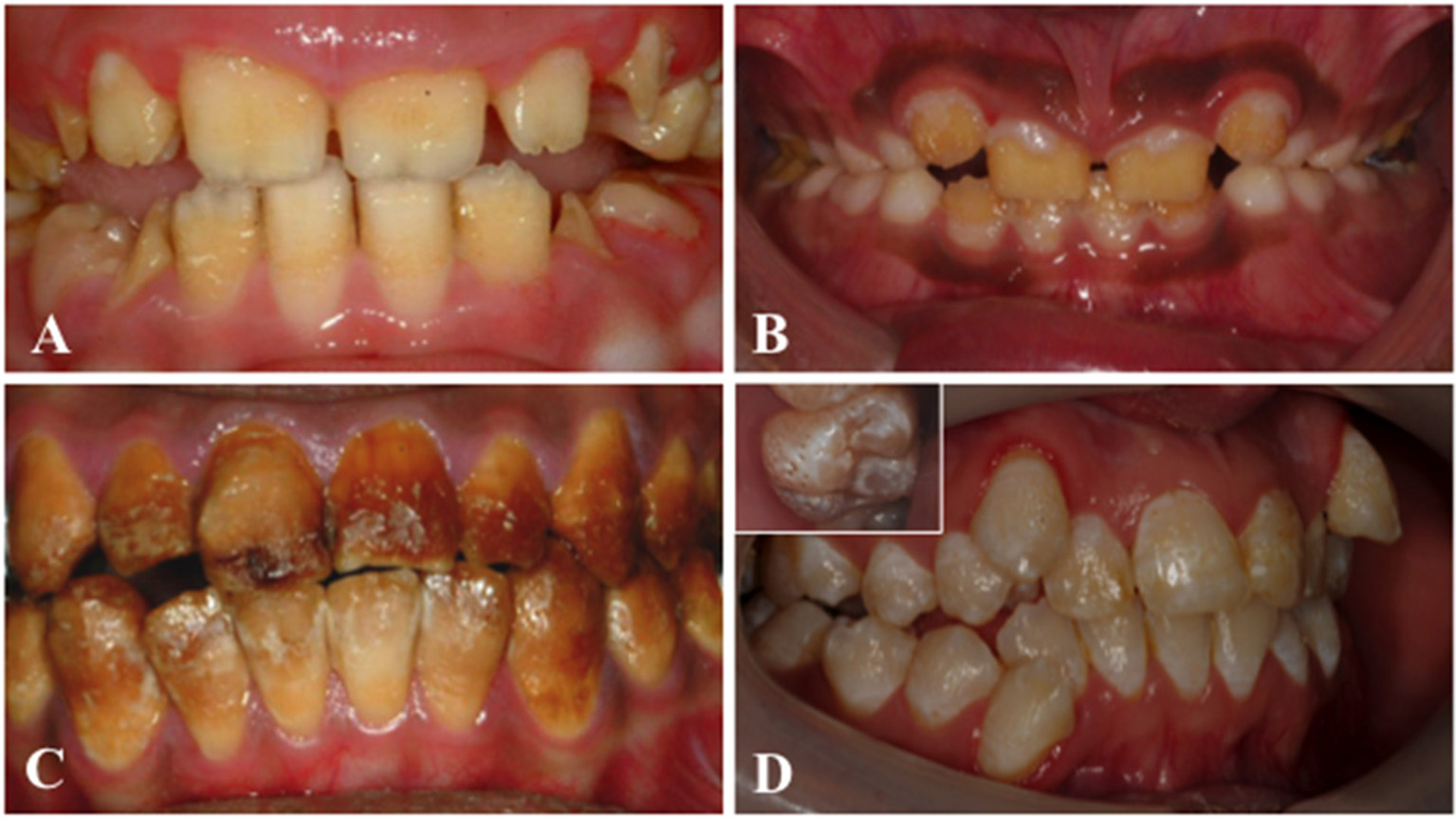
Image taken from ‘Amelogenesis imperfecta: Genes, proteins, and pathways‘ (see references list below). Reproduced according to the terms of the CC-BY 4.0 license.
Radiographic features include enamel that is either thinner or absent (hypoplastic phenotype), or a lack of differential radiodensity when comparing dentine and enamel (this is seen in the hypocalcified and hypomaturation phenotypes). These enamel changes can be visible on the unerupted dentition and can help with phenotype diagnosis. They can be associated with taurodontism on radiographic examination.
There are also multiple syndromes in which AI is part of the phenotype (this is called syndromic AI). It’s important that teeth are assessed by a specialist dentist, as tooth phenotypes can be incorrectly assumed to be a result of poor hygiene.
Genomics
AI is genetically heterogeneous, with multiple inheritance patterns. Variants in over 90 genes are known to cause it either as an isolated condition or as part of a syndrome. Pathogenic variants in COL17A1, MMP20, FAM83H, ENAM and AMELX are most commonly identified in individuals presenting with isolated AI. Variants in FAM20A are most commonly identified in individuals presenting with syndromic AI.
Diagnosis
AI is typically diagnosed in childhood. Diagnosis is usually based on clinical and radiographic features as listed above. The most common classification used is Witkop, 1988 (see the references list below), with hypoplastic, hypomaturation and hypocalcified categories as distinct types, alongside mixed phenotypes and syndromic cases. Modes of inheritance and causative genes have now been linked to each phenotype.
For many years, AI was not confirmed through DNA sequencing. However, given its genetic heterogeneity and the syndromic nature of some forms of AI, molecular confirmation now facilitates identification of future potential health concerns and, in some cases, prompts their prophylactic treatment.
For information about genomic testing, see ‘Child with yellow/brown teeth that are “crumbly“‘.
Inheritance and genomic counselling
Prevalence of AI varies, with studies reporting 1 in 4,000 people in Sweden being affected and 1 in 14,000 people in the United States.
Delivery of results can initially be conducted by the requesting dentist, with additional support from clinical genetics in certain cases – for example, where there is a link with a separate medical condition, such as enamel renal syndrome, or when familial testing is indicated. Genetic counselling in AI depends on the mode of inheritance and gene variant detected, as over 90 causative genes have been identified in relation to AI. Over 30 of these are currently included in the diagnostic R340 panel within the National Genomic Test Directory.
Autosomal dominant, autosomal recessive and X-linked inheritance can occur, as well as new (de novo) genetic changes, in which the individual’s parents are unaffected.
Autosomal dominant inheritance
The relative frequencies of these inheritance patterns vary; however, some of the commonly identified genetic variants cause disease with autosomal dominant inheritance (variants in the COL17A1, FAM83H and ENAM genes).
- Individuals affected by an autosomal dominant condition have one working copy of the gene, and one with a pathogenic variant.
- The chance of a child inheriting the gene with the variant from an affected parent is 1 in 2 (50%). The same is also true of genetic variants that arise de novo.
- Incomplete penetrance, in which not everyone who has the variant develops the disease, has not been reported for AI but may exist.
Autosomal recessive inheritance
Pathogenic variants in MMP20 cause autosomal recessive AI.
- If both parents are carriers of an autosomal recessive condition, with each pregnancy there is a:
- 1-in-4 (25%) chance of a child inheriting both gene copies with the pathogenic variant and therefore being affected;
- 1-in-2 (50%) chance of a child inheriting one copy of the gene with the pathogenic variant and one normal copy, and therefore being a healthy carrier themselves; and
- 1-in-4 (25%) chance of a child inheriting both normal copies and being neither affected nor a carrier.
X-linked inheritance
The least common inheritance pattern is X-linked. The only gene associated with X-linked isolated AI is AMELX. Notably, females can have stripes of good- and bad-quality enamel as a result of being a heterozygote for a pathogenic AMELX variant.
- X-linked recessive conditions are usually only present in males.
- Males with X-linked conditions cannot pass the variant on to their sons, but they always pass their affected X chromosome to their daughters. If the condition is recessive, their daughters will be carriers for the condition.
- Female carriers of X-linked recessive conditions have a second, working copy of the gene and are therefore usually unaffected, or affected only mildly.
- Sons of female carriers of X-linked recessive conditions have a 1-in-2 (50%) chance of being affected by the condition, and their daughters have a 1-in-2 (50%) chance of being carriers.
- X-linked dominant conditions can affect both males and females.
- Affected males tend to have more significant disease than affected females.
If you are discussing genomics concepts with your patients, you may find it helpful to use the visual communication aids for genomics conversations.
Management
Management of AI is case-dependent and requires specialist dental input. Overall, there are two aims:
- maintain tooth structure and therefore function; and
- improve aesthetics.
However, evidence-based treatments for AI are lacking.
Specific challenges include:
- masking discolouration of teeth;
- hypersensitivity during treatment;
- poor oral hygiene (due to pain upon brushing and/or flossing);
- loss of inter-occlusal space due to tooth loss or wear; and
- decreased bond strength in the case of restorative treatments.
Treatment options vary, depending on patient factors such as age and dental anxiety. Management of AI in childhood is important, but challenges of care also extend into adulthood. Protecting vulnerable enamel (due to hypoplastic or hypocalcified phenotypes) is key. Treatment options can extend from minimally invasive fissure sealants, to direct composite restorations, to more advanced, indirect options, such as onlays or crowns.
If a genetic diagnosis is made, a multidisciplinary team may be required to manage treatment. For example, if pathogenic variants within FAM20A are identified, regular monitoring of kidney health and preventive treatment with medications that reduce the risk of kidney calcification may be recommended.
No genetic therapies have been developed for the prevention of AI.
Resources
For clinicians
- Genomics England: NHS Genomic Medicine Service (GMS) Signed Off Panels Resource: Amelogenesis imperfecta
- National Organization for Rare Disorders: Amelogenesis imperfecta
- NHS England: National Genomic Test Directory
- OMIM: Phenotypic series PS104500 Amelogenesis imperfecta
References:
- Bloch-Zupan A, Rey T, Jimenez-Armijo A and others. ‘Amelogenesis imperfecta: Next-generation sequencing sheds light on Witkop’s classification‘. Frontiers in Physiology 2023: volume 14. DOI: 10.3389/fphys.2023.1130175
- Lakhani S, Monteiro J, Agel M and others. ‘A UK-based consensus on clinical decision flowcharts for managing childhood amelogenesis imperfecta in the permanent dentition‘. European Archives of Paediatric Dentistry 2026: volume 27, pages 135–145. DOI: 10.1007/s40368-025-01127-1
- Smith CEL, Poulter JA, Antanaviciute A and others. ‘Amelogenesis imperfecta: Genes, proteins, and pathways‘. Frontiers in Physiology 2017: volume 8. DOI: 10.3389/fphys.2017.00435https://onlinelibrary.wiley.com/doi/10.1111/j.1600-0714.1988.tb01332.x
- Witkop CJ Jr. ‘Amelogenesis imperfecta, dentinogenesis imperfecta and dentin dysplasia revisited: Problems in classification‘. Journal of Oral Pathology & Medicine 1988: volume 17, issue 9–10, pages 547–553. DOI: 10.1111/j.1600-0714.1988.tb01332.x